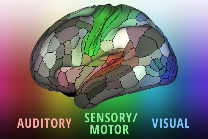
A brain map that includes more than doubled the number of distinct areas known in the human cortex, from 83 to 180, has been published. It combines data from four imaging technologies to bring high-definition to brain scanning. Washington University’s Matthew Glasser and David Van Essen led the global research team.
1200 young adult brains were scanned using MRI, which reveals the structure of the brain; fMRI, which registers brain activity while resting; task-based fMRI, which registers activity while engaged in mental exercises; and diffusion imaging, which reveals the paths of neurons.
By aligning the brain areas using the combined scanning protocol, an extraordinary degree of precision was achieved.
Potential applications include identifying biological markers for neurological diseases and mental illnesses, and the ability for neurosurgeons to better define tissue.
Click to view Nature video
Join ApplySci on February 7-8 at Stanford University for Digital Health + NeuroTech Silicon Valley 2017